In one of my first posts, I have talked about various human systems such as the circulatory system and respiratory system. The digestive system was one of them, and today I will be going over this system in more detail. We will cover digestion and absorption in the small intestine.
Digestion in the Small Intestine
During the process of digestion, food is broken down into monomers so that the villi can easily absorb them and the lymph or the blood can take them up. After large food molecules are broken down into small pieces (mechanical digestion), the wall of the intestine and the pancreas produce enzymes needed for chemical digestion, which is the breaking down of small food units into monomers.
Two types of muscle contraction, both of which are peristalsis, pass along the intestine. The contraction of circular muscle prevents food from being pushed back towards the mouth. The contraction of longitudinal muscle moves the food along the gut. When both layers of the muscle contract, food is mixed with enzymes in the small intestine. Examples of enzymes secreted by the pancreas include lipase, endopeptidase, and amylase. Lipase breaks down lipids (fats and oils) into fatty acids and glycerol. Endopeptidase breaks down polypeptides into shorter peptides. Amylase breaks down starch into maltose.
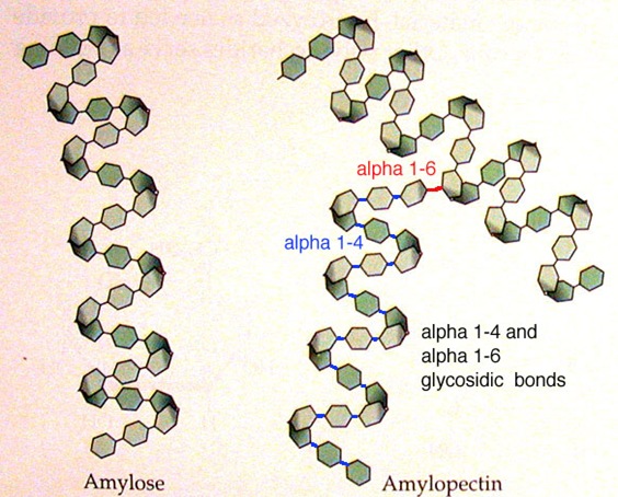
There is actually much more to the last example: digestion of starch. Amylose and amylopectin are two types of molecule in starch. They are both polymers of alpha-glucose linked by 1,4 bonds. But in amylose, the chains are unbranched whereas in amylopectin, the molecule is branched by 1,6 bonds. These 1,6 bonds cannot be broken down by amylase, and fragments of the amylopectin molecule that is not digested are called dextrins. Dextrins are then digested by dextrinase in the membranes of microvilli.
Absorption
After food is digested in the stomach and small intestine, they are absorbed by intestinal villi. Substances are taken in by cells and the blood to supply nutrition. Due to the variety of molecules that needs to be absorbed, there are a number of different methods by which the small intestine takes in molecules. Simple diffusion (nutrients pass down the concentration gradient) takes in hydrophobic nutrients like fatty acids and monoglycerides. Facilitated diffusion (nutrients pass down the concentration gradient through specific channel proteins) takes in hydrophilic nutrients such as fructose. Active transport (nutrients travel against the concentration gradient) absorbs mineral ions like iron, calcium, and sodium. Endocytosis (small droplets of fluid are passed by means of vesicles) absorbs triglycerides and cholesterol.
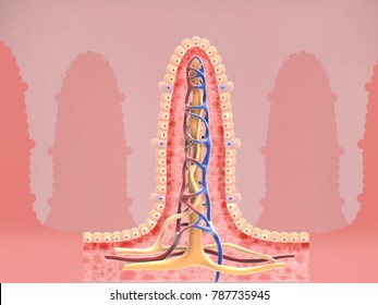
In the human digestive system, nutrients are absorbed by the epithelium – a single layer of cells forming the inner lining of the mucosa. The rate of absorption, therefore, is largely affected by the surface area of the epithelium. This area is increased by villi and microvilli. Villi are small projections of the mucosa, and it is between 0.5 and 1.5 mm long. They increase the surface area by approximately a factor of 10. They take in minerals ions and vitamins digested in the duodenum of the small intestine.
Modelling Absorption
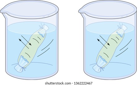
Absorption by the epithelium can be modeled with dialysis tubing. Though there are many ways to design this apparatus, I will present an example using Cola. Cola contains a mixture of substances that represents digested and undigested substances in the body. Water will be poured outside the bag to see which substances diffused through. The results were that glucose and phosphoric acid, which are both small-sized molecules, diffused through the tubing but larger polymers of sugar could not.
Works Cited
Allott, Andrew. Biology: Course Companion. Oxford University Press, 2014.
Kochunni, Deena T, and Jazir Haneef. “Difference between Amylose and Amylopectin.” Difference between Amylose and Amylopectin, http://www.majordifferences.com/2013/02/difference-between-amylose-and_17.html#.XbEZwJMzYUs.
Shutterstock. “Dialysis Tubing Images.” Shutterstock, http://www.shutterstock.com/search/dialysis+tubing.
Shutterstock. “Intestinal Villi Images.” Shutterstock, http://www.shutterstock.com/search/intestinal+villi.


Leave a comment